Source:
http://www.hospimedica.com/critical_care/articles/294732895/new_endoscopic_device_detects_esophageal_cancer.html
HospiMedica International staff writers
Posted on 12 Jan 2011
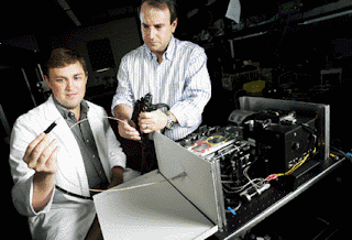
Image: Biomedical engineers Neil Terry (left) and Adam Wax look over the endoscope a/LCI device (photo courtesy Duke Photography).
An innovative device holds the promise of being a less invasive method for testing patients suspected of having Barrett's esophagus (BE), a change in the lining of the esophagus due to acid reflux.
Researchers at Duke University (Durham, NC, USA) developed the new device, which is based on an endoscope with a miniature light source and sensors at its end. Physicians shine short bursts from the light source at locations of suspected disease, and the sensors capture and analyze the light as it is reflected back. The devices is based on angle-resolved low coherence interferometry (a/LCI), a technique capable of separating unique patterns of the cell nucleus from the other parts of the cell, and provide representations of its changes in shape in real time. By detecting characteristic changes within the layer of the epithelial lining of the esophagus, BE can be detected and treated earlier.
A preliminary clinical trial of the device was conducted at the University of North Carolina (UNC; Chapel Hill, USA) on 46 patients with BE. The researchers measured the mean diameter and refractive index of cell nuclei in esophageal epithelium at 172 biopsy sites. At each site, an a/LCI measurement was correlated with a concurrent endoscopic biopsy specimen, which was assessed histologically and classified as normal, nondysplastic BE, indeterminate for dysplasia, low-grade dysplasia (LGD), or high-grade dysplasia (HGD). The researchers found that the a/LCI nuclear size measurements separated dysplastic from nondysplastic tissue at a statistically significant level for the tissue segment 200 μm to 300 μm beneath the surface, with an accuracy of 86%. The results were published in the January 2011 issue of Gastroenterology.
“Currently, we take many random tissue samples from areas we where we think abnormal cells may be located,” said lead author UNC gastroenterologist Nicholas Shaheen, MD. “This new system may make our biopsies smarter and more targeted. Early detection is crucial, because the cure rate for esophageal cancer that is caught early is quite high, while the cure rate for advanced disease is dismal, with a 15% survival rate after five years.”
“By interpreting the way the light scatters after we shine it at a location on the tissue surface, we can the spot the tell-tales signs of cells that are changing from their healthy, normal state to those that may become cancerous,” added Neil Terry, a PhD student working in the laboratory of Adam Wax, PhD, an associate professor of biomedical engineering at Duke, who developed the device. “Specifically, the nuclei of precancerous cells are larger than typical cell nuclei, and the light scatters back from them in a characteristic manner. When we compared the findings from our system with an actual review by pathologists, we found they correlated in 86% of the samples.”


No comments:
Post a Comment